


The deformation of Valgus of the feet is often congenital. However, in some cases - with paralysis, traumatic lesions - they can already appear in the mature period of life. The main symptoms of the pathology are the gaze in the area of the feet and muscles of the lower part of the leg, a visible violation of the shape of the feet and a change in the pace. The diagnosis of the disease is carried out using a clinical examination, an X -ray, electromyography, etc. It includes conservative and surgical methods. However, correct effectiveness is observed only in reconstructive operations.
What is this disorder?
Valgus deformation is the curvature of the foot, characterized by the flattening of its longitudinal arch. Generally, the internal edge of the foot is lowered ("drops") and the heel turns out.
The foot of a person, by virtue of his position, assumes the pressure of the entire mass of the human body. For this reason, it has a special anatomical structure, which allows amortization, balance and stabilization movements. However, an important component for the implementation of these tasks is the correct form of arrest.
Today, the most important problem of traumatology and orthopedics is the deformation of the foot. The meeting is estimated at 30-58%, where 2/3 of cases constitute congenital disorders.
The pathology is largely socially significant, because it covers all the age groups of the population and also helps to curve the spinal column, the early development of osteochondosis and the arthrosis of the joints of the lower ends.
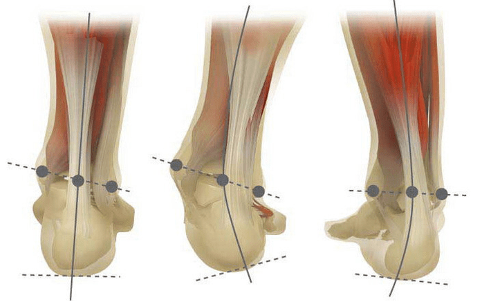
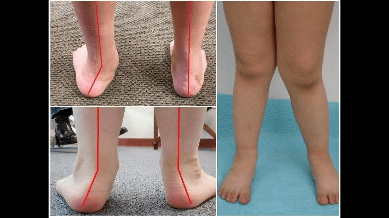
When you get your feet (if you look at them from behind), a deformation similar to x at the ankle level is formed: the ankles are in contact, while the heels are at a distance of 5-6 centimeters from each other.
Very often, the pathology is of a congenital nature and is diagnosed in children in the hospital (or immediately after the start of the walk). A similar condition is regulated up to 5 years, after which (in the absence of adequate treatment), a child develops flat feet.
Why does it stand?
It is believed that the main reason for the appearance of the deformation of the valgus of the foot is an inadequate function of the rear tibial muscle or weakness of the binding apparatus.
Today other factors are distinguished in the development of the pathology:
- Congenital anomalies with the incorrect position of the bones of the feet or shortening of the tendons (vertical bone of ram, tendon of the short pile);
- Posture disorders when the deformation of the feet compensates the curvature of the spinal column;
- Traumatic lesions (fractures of the feet bones, lower legs, hips or knee, breakage of the binding and tendon apparatus);
- Paralysis (immobilization) due to damage to the nervous system for encephalitis, polio, stroke, violation of cerebrospalous melts for hernias, etc. ;
- Spasm (constant contraction) of the muscles of the lower part of the leg;
- Concomitant diseases: pathology of the bone system with vitamin D (rachit), diabetes mellitus, osteoporosis (reduction of bone density), thyroid gland and compromised paratyroid, etc. ;
- Increase in body weight, including rapid weight gain in menopause or during pregnancy.
The development of the pathology is also facilitated by incorrectly selected shoes or an excessive correction of the club's foot during childhood.
Grade and Stadium of the disease
The severity of the pathology (power of the event) is divided by degrees:
- Light with a height of the vault of 1, 5-2 centimeters and an angle of inclination of the heel at 15 degrees;
- The average, when the arch is flattened in the 1st centimeter and the angle decreases at 10 degrees;
- Height at the height of the vault up to 0, 5 cm and the angle of inclination of the heel is 5 degrees.
Depending on the involvement of some structures, the following phases of the curvature are distinguished:
- There are no bone deformations, the pain is determined on the internal surface of the ankle (in the area of the fixing of the rear tibial muscle);
- The curvature is light, the heel is slightly rejected;
- The foot is assigned and the deformation is fixed (not correctly correct);
- Wending is observed not only in the foot, but also in the artery joint.
Symptoms
In the first phase, patients are disturbed by periodic pain after long walks or long vertical loads (standing or sitting on the foot). As a rule, pain syndrome intensifies when walking in shoes selected improperly. The next phase of the disease is associated with the occurrence of the curvature of the foot: patients in the erect position are not based on the outer edge of the foot, but with all its area. A slight change in the pace is observed.
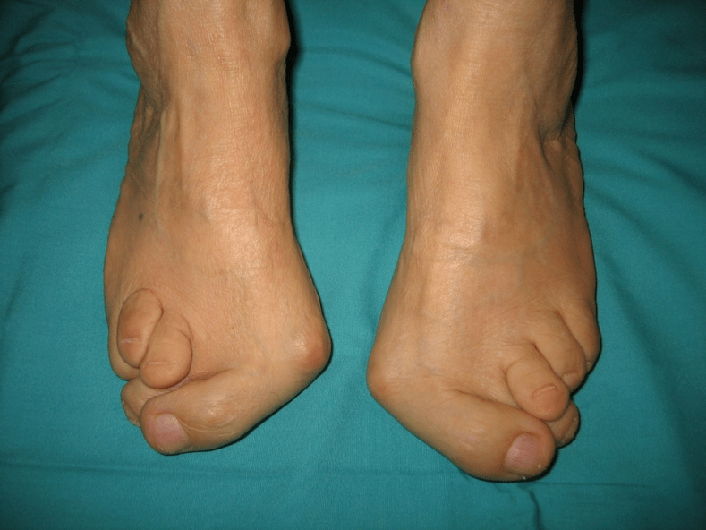
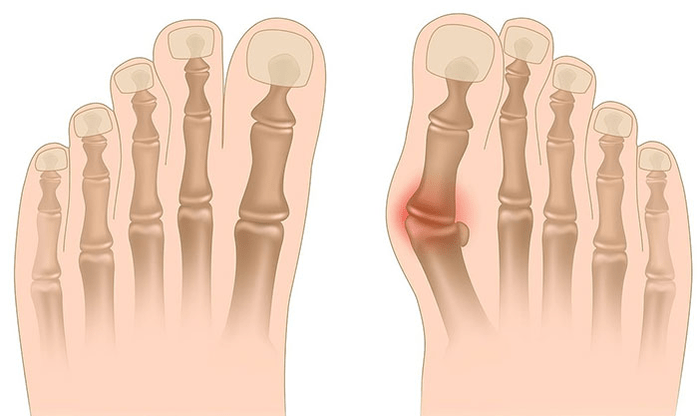
In the third phase, the protruding bone protruding is determined (considerably lower than the ankle on the internal surface of the ankle), as well as a strong deviation of the heel outside (the patient is based on the inner edge of the heel bone). The advanced deformation of Valgus dei Pieda is characterized by a pronounced curvature both of the foot itself and of the artery joint. Patients complain of severe pain in the muscles of the lower part of the leg, as well as a significant violation of the pace: the knees rub each other, while the right and left foot is located at a certain distance.
The serious curvature of the feet is often complicated by the deformation of the spine (scoliosis with different positions of the shoulders and the wings of the pelvis), of the osteochondosis (damage to the intervertebral disc with the formation of an hernia) or arthrosis (premature wear of intraaricular cartils in the lower leg and a tank top).
How to diagnose?
The diagnosis of the curvature of the foot consists of:
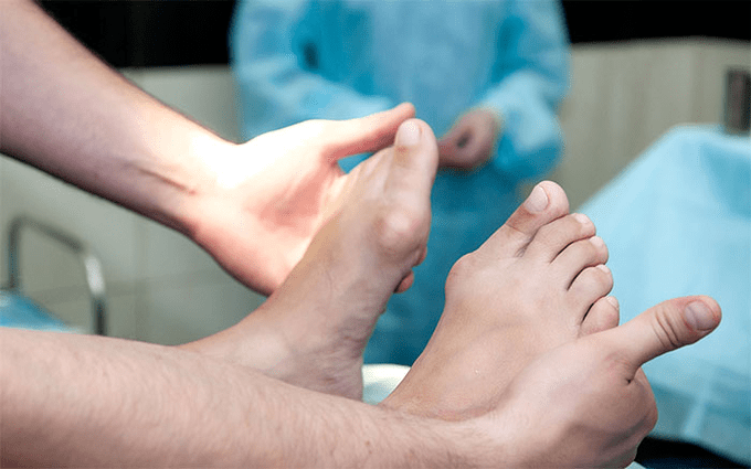
- The clinical inspection, during which the orthopedist detects a decrease in the arches of the foot, the deviation of the bones of the heel and the RAM, the visible "disappearance" of the outside and the protrusion of the internal ankles.
- X -Ray - An economic and information method with which it is possible to determine the change in the corners of the bone inclination and the linear parameters of their relationship. These indicators are necessary to make a final diagnosis and clarify the degree of deformation.
- The method of recording the phases aimed at determining the exact functional state of the limb. The method consists in the recording of the time of the support of the individual parts of the foot during the execution of a passage. During the study, the rolling phases are also studied, which reflect the balance of the lower limbs muscles.
- Dynamic electromyography, which records the electrical activity of the muscles studied and its dependence on the step of the step.
- Photoplesmography with digital processing that allows you to obtain all standard indicators and determine the type/degree of curvature with high precision.
A further consultation of a neurologist (with deformations due to spasms or paralysis), endocrinologist (in the case of diabetes or disturbances of the thyroid/wallpaper gland) and gynecologist (when the threatening) can be requested. If the curvature of the foot appeared in the background of osteoporosis, densitometry is necessary: the study of bone density.
Treatment
Among the main methods for the treatment of the curvature of Valgus of the feet, conservative and operational are distinguished. Do not destroy the painful joints with ointments and injects!
Conservative approach
This type of help aims to get rid of the symptoms of the disease, but does not eliminate the main cause of the pathology.
The technique includes:
- the use of orthopedic sunspasses to support the bone with the plus, the arch of the foot, as well as the elimination of the Valgus lesions of the middle and the back of the foot;
- Tasso: fix the foot and ankle using special adhesive ribbons with adequate elasticity. The tape ribbon is worn around the clock within 3-5 days, after which they are replaced;
- sew orthopedic shoes according to individual standards;
- The use of orthosis and other fixing devices on the foot and ankle.
Conservative methods also include physiotherapy procedures (Ozokrete, paraffin applications, electrophoresis, magnetic effects), massage and a complex of physiotherapy exercises, developed for a specific clinical case. Be careful! Today, most experts prefer surgical treatment methods, since conservative therapy is ineffective (according to statistics, it is useless in 60% of cases).
Surgery
The volume of operating and its type depend on the direct phase of the disease. Therefore, the first degree of valgus deformation is treated by syvectomy (removal of the tendon shell for the correction of the general voltage) or the osteotomy (dissection) of the heel to return to the anatomically correct position. In the second phase of the development of the disease, the curves of the finger tendon is used. This intervention is usually performed against the background of the dissection of the heel or an arthrodesis to RAM (surgical immobilization of the joint between the RAM and the dash bones).
The curvature of Grado III requires an arthrodesis of different joints of the foot simultaneously: the most devoid of cup-cubic and designated by RAM. This three -mature immobilization is often completed by the dissection of the heel bone. In phase IV of the pathology, reconstructive operations are needed not only on the foot, but also on the ankle. In this case, the instability of the binding apparatus is regulated by transplants (from one's body or artificial materials). The volume of the operations on the foot itself is the same as the grade III of curvature.
Recovery period
Rehabilitation includes walking without support on the leg managed for 2 months. At the same time, the patient must wear the fault of removable chalk from 1, 5 to 3 months. Active movements are recommended in the stock foot after 1, 5 months after surgery. Within the 3rd month, a complex of strengthening of physical education is introduced. However, active sports and sports activities are prohibited. It is worth noting that it is possible to judge the final result of surgery only six months later.
Preventive measures
The prevention of the Valgus dell'Artesto deformation includes the following measures:
- Early correction of congenital anomalies with the improper arrangement of the bones of the feet or the shortening of the tendon beans (vertical defrosted bone, tendon of the short heel);
- Correction of posture disorders (scoliosis, etc. );
- Timely treatment of traumatic lesions (fractures of the feet bones, lower part of the leg, thigh or knee joint, breakage of the binding apparatus and tendon);
- The correct rehabilitation after paralysis (immobilization) due to the damage to the nervous system for encephalitis, polio, stroke, violation of cerebrospalous roots for hernias, etc. ;
- Spasm relief (constant reduction) of the muscles of the lower part of the leg;
- Therapy for concomitant diseases: pathologies of the bone system with vitamin D (rachit), diabetes mellitus, osteoporosis (reduction of bone density), compromised functionality of the thyroid and wallpaper glands, etc. ;
- A decrease in body weight to normal (especially with a rapid weight gain in postmenopausal or due to pregnancy);
- Selection of orthopedic shoes or the use of supinators;
- Moderate correction of the foot club without treatment with excess of "hyper-correction" which leads to an excretion of the secondary valgus of the feet.
The prevention of the progression of the disease consists in using conservative methods and early reconstructive operations. In this case, physical activity is limited in order to prevent the destruction and curvature of the ankle joints. Remember, the timely treatment of the deformation of Valgus of the feet not only improves the quality of life of patients, but also prevents the development of osteochondrosis and arthrosis of the knee or hip joints!